|
A thirty-five year old white male was admitted to the hospital for pleuritic chest pain and fever in the fall of 2009.
A lifelong resident of Oregon who had suffered with crohn's disease since childhood, he had become ill with headache, muscle aches, and fever approximately two weeks prior to hospital admission. Approximately a week prior to admission he had also developed pleuritic left sided chest pain for which he sought medical attention. Although he lacked a significant cough, his chest x-ray suggested pneumonia, and he received outpatient therapy with azithromycin with subsequent addition of levofloxacin when chest pain and fevers (to 103o) persisted.
His treatment for crohn's disease was with mesalamine and azathioprim. He worked in an office and did not typically engage in outdoor pursuits. He home was in an urban environment in a moderately sized community in the Willamette Valley. Approximately three months prior to his illness he and his family had visited southern California traveling through the Central Valley en route. His wife and two young children had not been recently ill. His wife was foreign born from a region where tuberculosis was of high prevalence, and there was concern about possible past exposures to persons with active turberculosis.
He was admitted to the hospital because of failure to improve. At the time of his admission he was described as in no acute distress, with lungs clear to auscultation, and oxygen saturation 98% on room air. Admission laboratories included WBC 8.4, Hgb 13.1 and normal LFTs. He was begun on broad spectrum antibiotic therapy with piperacillin/tazobactam and vancomycin, and azathioprim was discontinued.
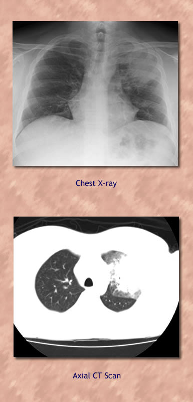
Laboratory investigation included CXR and chest CT scan (at left). Urine legionella antigen, urine pneumococcal antigen, serum cryptocococcal antigen, and influenza rapid tests were negative. Blood cultures and sputum AFB smears were also negative. Largely because of concern about the possibility of tuberculosis, the patient underwent bronchoscopy with LUL bronchoalveolar lavage. The bronchoscopist described slightly erythematous uppper airways without purulence or endobronchial abnormality.
The patient appeared to improve on the broad spectrum antibiotic therapy and was discharged on cefdinir after a five day hospitalization.
|