Infectious Diseases Case of the Month #5 |
|||
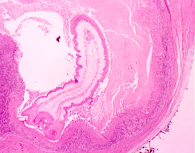 |
A 17 y.o. hispanic male was seen in infectious diseases clinic at the request of his primary physician because of a subcutaneous parasitic infection. Born near Acapulco, Mexico the patient had moved to Chelan, Washington (North Central Washington) with this family when he was 12 years old. He was in good health and was a high school student. He liked to swim in Lake Chelan, a large mountain lake, and occasionally the Columbia River. His family had dogs when they lived in Mexico but not during their five year residence in Washington State. He liked seafood and occasionally his family would butcher a pig for their own consumption. He had not traveled out of North America. His primary physician had excised a small (2-3 cm) subcutaneous lump from his left anterior chest that had developed spontaneously and asymptomatically a couple weeks previously. The patient felt well. A photomicrograph of the excised cystic structure is at left. Click here for a close up view of the anterior portion of the parasite. |
||
What organism is most likely to have caused the development of this subcutaneous cyst? |
|||
 |
The previous images were of a juvenile cestode, most likely Taenia solium, already evaginated from a dermal cysticercus. This case is a very difficult one to definitively diagnose for non-parasitologists unskilled in the microscopy of parasitic diseases. The interpretation of the specimen was performed by pathologists at the Armed Forces Institute of Pathology (AFIP). Cysticerci of Taenia solium can be found in multiple tissues such as in the subcutaneous tissues in the case described. Their occurrence is generally benign. The exception is cysticerci in the central nervous system (neurocysticercosis) where their presence can cause seizures; neurocysticercosis is the most common cause of focal motor seizures in certain areas of the world where the disease is endemic. The life cycles of Taenia solium and Taenia saginata are pictured at left. Humans are the definitive host for both species of tapeworm. Though not illustrated in the diagram, humans can also be an intermediate host for Taenia solium where the larvae are encysted in tissue (cysticercosis as described above). The scoleces of the respective species are also pictured at left where the resemblance to the case photomicrograph is evident (notice that the scolex of Taenia saginata lacks the central hooks). Most of the other parasites in the previous list (see choices in original format) can also have cutaneous/subcutaneous manifestations. Infection with Drancunulus medinensis (Guinea worm) manifests as a papule typically on the lower extremities which blisters and ulcerates as the female worm releases her eggs. Infection with Leishmania mexicana (New World cutaneous leishmanias) can cause a variety of skin lesions ranging from ulcerative to eruptive/exudative. Subcutaneous nodules are a hallmark of infection with Onchocercus volvulus (river blindness). Echinococcus granulosus infection (hydatid cyst disease) is not typically manifested by skin findings; nonetheless, it like Taenia solium, is a cestode (tapeworm) for which humans may be an accidental intermediate host. Ref: Peters, W., Gilles, H.M., Tropical Medicine and Parasitology, 4th ed., Mosby-Wolf, 1995. |
||
| Home Case of the Month ID Case Archive | Your Comments/Feedback | ||